All published articles of this journal are available on ScienceDirect.

A Retrospective Analysis of Removable Partial Denture Design in Partially Edentulous Patients Attending a Dental Teaching Hospital
 iD
Arwa U. Alsaggaf1
iD
Arwa U. Alsaggaf1
Authors Info & Affiliations
Abstract
Introduction/Objective
Removable Partial Dentures (RPDs) remain a widely used treatment modality for the rehabilitation of partially edentulous patients. Understanding the distribution of Kennedy classifications and the patterns of RPD design is essential for improving treatment planning and dental education. This study aimed to retrospectively analyze RPD design characteristics among partially edentulous patients attending a dental teaching hospital.
Methods
A retrospective cross-sectional study was conducted using patient records from October 2015 to April 2022. A total of 551 partially edentulous patients aged over 20 years were included. Demographic data, Kennedy classifications, Direct retainers, and Major connector designs were collected and analyzed. Descriptive statistics and chi-square tests were performed using STATA version 14.2, with statistical significance set at p < 0.05.
Results
Among the 551 patients, 67.5% were female, and 32.5% were male, with a mean age ranging from 43.4 ± 12.6 years. Kennedy Class III was the most prevalent classification overall, particularly in the maxillary arch, whereas Class I was more frequently observed in the mandibular arch (p < 0.0001). The Akers’ clasp was the most commonly used direct retainer (45.6%), whereas the RPI clasp was predominantly associated with Kennedy Class I cases (64.6%). The mid-palatal strap was the most common maxillary major connector (59.8%), and the lingual bar was the most frequently used mandibular major connector (77.8%). Sex-disaggregated analyses showed a significant association between age groups and Kennedy classification in female patients (p = 0.007), but not in male patients (p = 0.06).
Discussion
The findings demonstrate distinct patterns in the distribution of Kennedy classifications and RPD design elements within the studied population. These patterns reflect established prosthodontic design principles and may assist clinicians and educators in optimizing RPD treatment planning and training.
Conclusion
Kennedy Class III was the predominant classification among partially edentulous patients, with Aker clasps, mid-palatal straps, and lingual bars representing the most frequently selected design components. The observed associations between arch type, classification, and design characteristics provide valuable information for evidence-based RPD design and dental education.
1. INTRODUCTION/ BACKGROUND
Partial edentulism remains a common oral-health problem that can impair mastication, appearance, speech, and oral-health-related quality of life. Studies conducted in Saudi Arabia have documented a substantial burden of missing teeth and partial edentulism across diverse adult populations [1–4].
Removable Partial Dentures (RPDs) remain an important treatment option because they are conservative, comparatively affordable, adaptable to changing clinical conditions, and suitable for patients who cannot receive fixed or implant-supported prosthesis [5, 6].
The success of RPD therapy depends on careful diagnosis and design. Appropriate selection of direct and indirect retainers, rests, denture bases, and major connectors is required to control movement, distribute functional loads, and protect abutment teeth and residual ridges [7–9]. Contemporary evidence also shows that technical complications-including loss of retention, framework or acrylic-base fractures, denture-tooth complications, and the need for relining or repair-remain clinically relevant [10].
RPD fabrication requires coordinated clinical and laboratory procedures, including surveying, design prescription, abutment preparation, impression making, framework fabrication, and clinical verification. Structured teaching and explicit design communication are therefore essential, while digital manufacturing methods are increasingly being evaluated for framework accuracy and reproducibility [11, 12].
Previous studies have reported considerable geographic variations in Kennedy classifications and RPD design patterns. However, fewer studies have evaluated whether designs produced in teaching institutions correspond to accepted biomechanical principles. Two recent investigations from Makkah and Umm Al-Qura University highlighted the importance of dentists’ design knowledge and the quality of student-generated RPD prescriptions [13, 14].
To the best of our knowledge, no prior study has jointly examined the distribution of partial edentulism and the corresponding RPD design patterns among patients treated at the Faculty of Dental Medicine, Umm Al-Qura University. Assessing these patterns is particularly relevant in an academic setting because student-generated designs reflect both clinical training standards and institutional supervision.
The study therefore aimed to determine the prevalence of Kennedy classes, identify the most frequently used components of the RPD framework, and evaluate associations with arch, age, and sex. Based on previous regional studies, the Kennedy Class III pattern was hypothesized to be the most prevalent one [3, 13, 14].
2. MATERIALS AND METHODS
This observational retrospective cross-sectional study included 551 patient records. Inclusion criteria required patients older than 20 years with partial edentulism. Records lacking key data or adequate documentation were excluded, and no data imputation was performed. Examiner calibration involved 30 records, with strong agreement (Kappa 0.95 intra-examiner and 0.90 inter-examiner, respectively). Data were analyzed using STATA version 14.2. Descriptive statistics and chi-square tests were applied, with significance set at p < 0.05. This study followed the Sex and Gender Equity in Research (SAGER) guidelines. The variable available in the archived clinical records was recorded as male or female and treated as biological sex for reporting and analysis; gender identity was not separately collected in the historical records.
The dental records of patients treated in the outpatient prosthodontics department between October 2015 and April 2022 were reviewed. Male and female patients aged over 20 years with partially edentulous maxillary, mandibular, or both arches were eligible. No participant was excluded on the basis of sex. Patients with completely edentulous arches and those with only missing third molars or supernumerary teeth were excluded. Records with incomplete documentation, missing RPD design sheets, or unclear photographic documentation were excluded to ensure data accuracy.
Patients presenting with shortened dental arches who were rehabilitated with RPDs were included, provided that the prosthetic replacement extended beyond functional premolar occlusion and fulfilled the inclusion criteria.
A total of 551 patient records that satisfied the inclusion criteria were analyzed. Demographic variables included age and recorded sex (male/female), along with marital status, occupation, and file number. Both sexes were included, and sex-disaggregated descriptive and inferential analyses were performed where applicable.
All patients were rehabilitated by undergraduate dental students under the direct supervision of prosthodontic staff members. For each case, the RPD design was determined following clinical and radiographic examinations. After abutment preparation, final impressions were obtained and submitted to the dental laboratory along with an approved design sheet for master cast fabrication and metallic framework construction. The clinical workflow included metallic framework try-in, teeth try-in, and final RPD delivery.
Clinical procedures were documented by students using digital cameras as part of their clinical requirement files. Standardized intra-oral photographs included maxillary occlusal, mandibular occlusal, and lateral views. Only records with clear and diagnostically acceptable images were included in the analysis. A representative image of each Kennedy classification is presented in Fig. (1).
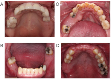
A representative image of each kennedy class. (A) kennedy class I. (B) kennedy class II. (C) kennedy class III. (D) kennedy class IV.
3. RESULTS
The sample included 372 female patients (67.5%) and 179 male patients (32.5%). The mean age was 43.4 ± 12.6 years. Kennedy Class III was the most common classification across both arches. Significant associations were found between the Kennedy classification and clasp types, as well as major connector selections. Sexdisaggregated distributions by age and Kennedy class are reported in Table 1.
| Age | Kennedy Classification | ||||||||||
|---|---|---|---|---|---|---|---|---|---|---|---|
| I | II | III | IV | Total | p-value | ||||||
| No. | % | No. | % | No. | % | No. | % | No. | % | ||
| Female | |||||||||||
| 20-29 | 20 | 25.3 | 16 | 20.3 | 41 | 51.9 | 2 | 2.5 | 79 | 100 | 0.007 |
| 30-39 | 22 | 29.3 | 23 | 30.7 | 30 | 40 | 0 | 0 | 75 | 100 | |
| 40-49 | 22 | 19.6 | 35 | 31.3 | 45 | 40.2 | 10 | 8.9 | 112 | 100 | |
| 50-59 | 12 | 15.6 | 15 | 19.5 | 46 | 59.7 | 4 | 5.2 | 77 | 100 | |
| 60-69 | 2 | 10.5 | 3 | 15.8 | 10 | 52.6 | 4 | 21.1 | 19 | 100 | |
| 70-79 | 1 | 10 | 3 | 30 | 5 | 50 | 1 | 10 | 10 | 100 | |
| Total | 79 | 21.2 | 95 | 25.5 | 177 | 47.6 | 21 | 5.6 | 372 | 100 | |
| Male | |||||||||||
| 20-29 | 4 | 20 | 1 | 5 | 15 | 75 | 0 | 0 | 20 | 100 | 0. 06 |
| 30-39 | 8 | 22.2 | 11 | 30.6 | 16 | 44.4 | 1 | 2.8 | 36 | 100 | |
| 40-49 | 8 | 16.3 | 15 | 30.6 | 21 | 42.9 | 5 | 10.2 | 49 | 100 | |
| 50-59 | 8 | 15.7 | 17 | 33.3 | 24 | 47.1 | 2 | 3.9 | 51 | 100 | |
| 60-69 | 7 | 38.9 | 8 | 44.4 | 2 | 11.1 | 1 | 5.6 | 18 | 100 | |
| 70-79 | 1 | 20 | 1 | 20 | 2 | 40 | 1 | 20 | 5 | 100 | |
| Total | 36 | 20.1 | 53 | 29.6 | 80 | 44.7 | 10 | 5.6 | 179 | 100 | |
The age and gender distribution of the study population is illustrated in Fig. (2).
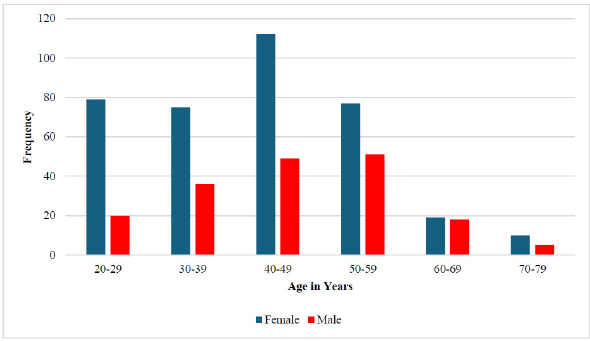
Age and gender distribution.
3.1. Kennedy Classification and Arch Distribution
A statistically significant association was observed between arch (maxilla vs mandible) and Kennedy classification (P < 0.0001) (Appendix Table 1).
Pairwise comparisons demonstrated that:
- Kennedy Class I was significantly more frequent in the mandible compared to the maxilla by 51.3% (95% CI: 33% – 70%; P < 0.0001).
- In the maxilla, Class III was significantly more frequent than:
- Class I by 43.6% (95% CI: 28.8% – 58.3%; P < 0.0001),
- Class II by 28.6% (95% CI: 14.8% – 42.5%; P = 0.0002),
- Class IV by 50% (95% CI: 34.8% – 65.1%; P = 0.0006).
A statistically significant association was also found between Kennedy classification and the presence of modification areas (P < 0.0001) (Appendix Table 2).
3.2. Kennedy Classification according to Age and Sex
Among female patients, a statistically significant difference was observed between age groups and Kennedy classifications (P = 0.007). Class III was the most prevalent classification across all female age groups, while Class IV consistently showed the lowest prevalence.
In male patients, Class III was the most prevalent classification across all age groups except the 60–69 age group, where Class II (44.4%), followed by Class I (38.9%), were more prevalent. Class IV showed the lowest prevalence in most male age groups, except in the 70–79 age group, where it was equally distributed (20%) with Classes I and II. No statistically significant difference was observed among male age groups (P = 0.06).
When comparing male and female patients:
- Classes I and II were generally more prevalent in females across most age groups, except in the 50–59 and 60–69 age groups, where males showed higher proportions (15.7% and 38.9%, respectively).
- Class III was more prevalent among males in the 20–29, 30–39, and 40–49 age groups (75%, 44.4%, and 42.9%, respectively), whereas females showed higher prevalence in older age groups.
- For Class IV, females demonstrated higher prevalence in the 30–39, 40–49, and 70–79 age groups (2.8%, 10.2%, and 20%, respectively).
Detailed sex-disaggregated distributions are presented in Table 1.
3.3. Association between Kennedy Classification and Clasp Type
A statistically significant association was identified between Kennedy classification and clasp type overall (P < 0.0001). The most frequently used clasp type was the Aker clasp (45.6%; 95% CI: 42.4% – 49%).
When analyzed by arch:
- In the maxilla, a significant association was observed (P < 0.0001), with the Aker clasp being the most common (42.6%; 95% CI: 37.8%–47.6%).
- In the mandible, a significant association was also found (P < 0.0001), with the Aker clasp again being the most frequent (48.1%; 95% CI: 43.6% – 52.6%) (Appendix Table 3).
3.4. Clasp Selection according to Kennedy Class
Regarding the most frequently used retainer for each classification:
- In Class III, the Aker clasp was most frequently used (61.3%).
- In Class II, the Aker clasp was also the most common (36%).
- In Class I, the RPI clasp was most frequently used (64.6%).
- In Class IV, the Double Aker clasp was the most prevalent (75.0%).
- RPA clasp was used only in Class I (11.8%) and Class II (5.7%) cases.
These distributions are illustrated in Fig. (3) and detailed in Appendix Tables 4 and 5.
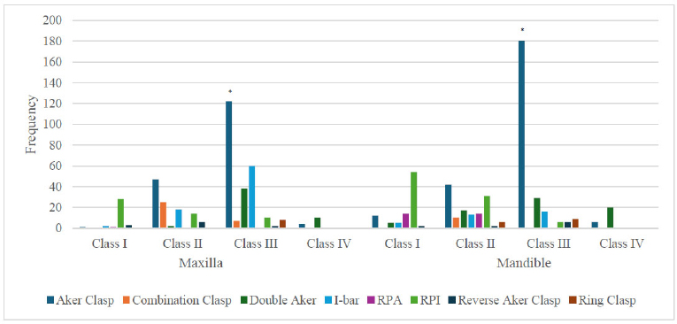
Different clasp types used for different maxillary and mandibular Kennedy classes.
3.5. Association between Kennedy Classification and Major Connector Type
In the maxillary arch, a statistically significant association was observed between Kennedy classification and major connector type (P < 0.0001). The Mid-Palatal Strap was the most frequently used major connector (59.8%).
In the mandibular arch, a statistically significant association was also found (P = 0.001), with the Lingual Bar being the predominant major connector (77.8%) (Appendix Table 6).
4. DISCUSSION
The predominance of Kennedy Class III is consistent with several regional studies and may reflect patterns of tooth loss, access to restorative care, and the age distribution of patients attending teaching hospitals. Because the study was retrospective and cross-sectional, causal explanations cannot be established.
The present study found Kennedy Class III to be the most frequent pattern. This agrees with reports from Pakistan and several Saudi populations [15–18], although other Saudi studies have found Kennedy Class I to be predominant [3]. These differences support the view that local demographic characteristics, referral patterns, and treatment settings influence the observed distribution.
The greater frequency of Class I in the mandible and Class III in the maxilla is broadly compatible with previously reported arch-related patterns [3, 17]. Class IV was the least frequent category in the present sample, a finding also reported in several epidemiological studies [3, 17].
Female patients represented 67.5% of the sample. A statistically significant age-related difference in Kennedy classification was detected among female patients, whereas the corresponding association among male patients did not reach statistical significance. These results should be interpreted with caution because the unequal sex distribution may reflect attendance patterns rather than a biological difference in tooth-loss patterns.
Aker clasps predominated in tooth-supported Class III cases, whereas RPI assemblies were most common in distal-extension Class I cases. This pattern is consistent with established biomechanical principles that favor circumferential retainers for bounded saddles and stress-releasing designs for tooth-tissue-supported prosthesis [7–9, 18].
The mid-palatal strap and lingual bar were the most frequently selected major connectors. Their use is compatible with the need for rigidity while minimizing unnecessary tissue coverage when anatomical conditions permit [7–9]. The findings suggest that most designs followed accepted principles, although the retrospective record-based approach did not permit direct assessment of clinical fit, patient comfort, or long-term performance.
Recent systematic reviews emphasize that RPD outcomes should be evaluated beyond framework design alone. Technical complications remain common enough to require planned maintenance [10], while digital and conventional workflows can both produce clinically acceptable frameworks, with differences in fit, efficiency, and patient-reported outcomes [12, 19, 20].
Patient satisfaction is influenced by retention, comfort, mastication, aesthetics, and adaptation to the prosthesis [21]. Periodontal status and maintenance are also important, as removable prostheses may increase risk in susceptible patients when plaque control and follow-up are inadequate [22].
Implant-assisted RPDs may improve support and patient-reported outcomes in selected distal-extension cases, but recent reviews indicate that evidence remains heterogeneous and that treatment design, attachment selection, and maintenance protocols require further investigation [6, 23].
Sex-related findings in this study should not be interpreted as evidence of intrinsic biological differences. The available records contained a binary male/female variable but did not capture gender identity or gender-related social determinants. Consequently, the results are generalizable primarily to patients attending this institution and should be confirmed in more representative multicenter samples.
Future studies should prospectively collect clearly defined sex and gender variables, recruit balanced samples when feasible, assess clinical outcomes and patient-reported measures, and evaluate whether design choices interact with age, sex, oral-health behavior, and socioeconomic factors.
5. STUDY LIMITATIONS
This study has several limitations that should be acknowledged. First, the retrospective design inherently limits control over data completeness and documentation accuracy. Although incomplete records were excluded and examiner calibration was performed, reliance on archived student-generated design sheets and photographic documentation may introduce variability in recording and interpretation of design.
Second, the study was conducted within a single academic institution, which may limit generalizability to other regions or private clinical settings. Institutional treatment protocols and supervision patterns may influence RPD design selection and may not fully reflect national practice trends.
Third, the analysis focused primarily on framework design patterns rather than clinical performance or long-term patient outcomes. Patient satisfaction, prosthesis longevity, and biological complications were not evaluated.
Additionally, although both acrylic and metallic RPDs were included in prevalence analysis, framework-specific design evaluation may differ between prosthesis types. The archived records used a binary male/female variable and did not document how sex was originally determined or collect gender identity; therefore, sex and gender-related interpretations are limited.
Future multicenter prospective studies incorporating clinical follow-up and patient-reported outcomes are recommended to further validate these findings.
CONCLUSION
Kennedy Class III was the most prevalent classification. RPD designs generally followed accepted prosthodontic principles, reflecting appropriate clinical training within the institution.
STUDY RECOMMENDATIONS
The findings of this study suggest that further research should be conducted in Makkah Al-Mukarramah using a representative sample size that covers all government and commercial dental clinics. Additionally, further studies should be conducted to estimate patient satisfaction with these RPDs, as it is a key objective of RPD construction.
STUDY CLINICAL SIGNIFICANCE
This valuable data will support oral health planners in suggesting plans to raise the service standards for oral health care in Saudi Arabia.
AUTHORS’ CONTRIBUTIONS
It is hereby acknowledged that all authors have accepted responsibility for the manuscript's content and consented to its submission. They have meticulously reviewed all results and unanimously approved the final version of the manuscript.
ETHICS APPROVAL AND CONSENT TO PARTICIPATE
Ethical approval was obtained from the Institutional Research Bioethics and Medical Committee, Umm Al-Qura University, Saudi Arabia (approval No. HAPO-02-K-012-2022-01-908).
HUMAN AND ANIMAL RIGHTS
All human research procedures followed were in accordance with the ethical standards of the committee responsible for human experimentation (institutional and national), and with the Helsinki Declaration of 1975, as revised in 2013.
CONSENT FOR PUBLICATION
All patient records were examined for informed consent signed by the patients, allowing the use of their information for dental faculty research.
AVAILABILITY OF DATA AND MATERIALS
Upon request, the corresponding author [A.M.] will make the datasets generated and analyzed during the current study available.
ACKNOWLEDGEMENTS
This study was fully self-funded by the authors.
APPENDIX
| - | Mandible | Maxilla | Total | |||||
|---|---|---|---|---|---|---|---|---|
| No. (%) | No. (%) | No. (%) | C.I. 95% | |||||
| Class I | 87 (15.8) | 28 (5.1) | 115 (20.9) | 17.7 - 24.5 | ||||
| Class II | 85 (15.4) | 63 (11.4) | 148 (26.9) | 23.3 – 30.7 | ||||
| Class III * | 127 (23.1) | 130 (23.6) | 257 (46.6) | 42.5 – 50.8 | ||||
| Class IV ** | 18 (3.3) | 13 (2.4) | 31 (5.6) | 4 – 7.9 | ||||
| Total | 317 (57.5) | 234 (42.5) | 551 (100) | |||||
** Significantly lower than other classes at 0.05 significance level.
| Modification Areas | Class I | Class II | Class III | Class IV | Total |
|---|---|---|---|---|---|
| No. | No. | No. | No. | No. (%) | |
| No | 51 | 21 | 40 | 19 | 131 (42) |
| Yes | 10 | 63 | 108 | 0 | 181 (58) |
| Clasp Type | Maxilla | Mandible | Overall | ||||||||||||
|---|---|---|---|---|---|---|---|---|---|---|---|---|---|---|---|
| Class I | Class II | Class III | Class IV | Total | Class I | Class II | Class III | Class IV | Total | Class I | Class II | Class III | Class IV | Total | |
| No. | No. | No. | No. | No. (%) | No. | No. | No. | No. | No. (%) | No. | No. | No. | No. | No. (%) | |
| Aker Clasp | 1 | 47 | 122 | 4 | 174 (42.6) | 12 | 42 | 180 | 6 | 240 (48.1) | 13 | 89 | 302 | 10 | 414 (45.6) |
| Combination Clasp | 0 | 25 | 7 | 0 | 32 (7.8) | 0 | 10 | 0 | 0 | 10 (2.0) | 0 | 35 | 7 | 0 | 42 (4.7) |
| Double Aker | 0 | 2 | 38 | 10 | 50 (12.3) | 5 | 17 | 29 | 20 | 71 (14.2) | 5 | 19 | 67 | 30 | 121 (13.3) |
| I-bar | 2 | 18 | 60 | 0 | 80 (19.6) | 5 | 13 | 16 | 0 | 34 (6.8) | 7 | 31 | 76 | 0 | 114 (12.6) |
| RPA | 1 | 0 | 0 | 0 | 1 (0.2) | 14 | 14 | 0 | 0 | 28 (5.6) | 15 | 14 | 0 | 0 | 29 (3.2) |
| RPI | 28 | 14 | 10 | 0 | 52 (12.7) | 54 | 31 | 6 | 0 | 91 (18.2) | 82 | 45 | 16 | 0 | 143 (15.8) |
| Reverse Aker Clasp | 3 | 6 | 2 | 0 | 11 (2.7) | 2 | 2 | 6 | 0 | 10 (2.0) | 5 | 8 | 8 | 0 | 21 (2.3) |
| Ring Clasp | 0 | 0 | 8 | 0 | 8 (2.0) | 0 | 6 | 9 | 0 | 15 (3.0) | 0 | 6 | 17 | 0 | 23 (2.5) |
| Total | 35 | 112 | 247 | 14 | 408 (100) | 92 | 135 | 246 | 26 | 499 (100) | 127 | 247 | 493 | 40 | 907 (100) |
Appendix Table 4.
| Clasp Type | Kennedy Classes | |||||||
|---|---|---|---|---|---|---|---|---|
| Class I | Class II | Class III | Class IV | |||||
| % | No. | % | No. | % | No. | % | No. | |
| Aker clasp | 10.20% | 13 | 36.00% | 89 | 61.30% | 302 | 25.00% | 10 |
| Combination clasp | 0.00% | 0 | 14.20% | 35 | 1.40% | 7 | 0.00% | 0 |
| Double Aker | 3.90% | 5 | 7.70% | 19 | 13.60% | 67 | 75.00% | 30 |
| I-bar | 5.50% | 7 | 12.60% | 31 | 15.40% | 76 | 0.00% | 0 |
| RPA | 11.80% | 15 | 5.70% | 14 | 0.00% | 0 | 0.00% | 0 |
| RPI | 64.60% | 82 | 18.20% | 45 | 3.20% | 16 | 0.00% | 0 |
| Reverse Aker | 3.90% | 5 | 3.20% | 8 | 1.60% | 8 | 0.00% | 0 |
| Ring | 0.00% | 0 | 2.40% | 6 | 3.40% | 17 | 0.00% | 0 |
| Total | 100.00% | 127 | 100.00% | 247 | 100.00% | 493 | 100.00% | 40 |
| Indirect Retainer | Kennedy Classification | - | ||||||||
|---|---|---|---|---|---|---|---|---|---|---|
| I | II | III | IV | Total | ||||||
| No. | % | No. | % | No. | % | No. | % | No. | % | |
| No | 0 | 0 | 0 | 0 | 148 | 47.3 | 19 | 6.1 | 167 | 53.4 |
| Yes | 61 | 19.5 | 85 | 27.2 | 0 | 0 | 0 | 0 | 146 | 46.6 |
| Total | 61 | 19.5 | 85 | 27.2 | 148 | 47.3 | 19 | 6.1 | 313 | 100 |
| Arch | Major Connector Type | Class I | Class II | Class III | Class IV | Total |
|---|---|---|---|---|---|---|
| No. | No. | No. | No. | No. (%) | ||
| Maxilla | Anterior Palatal Strap (U-shaped palatal strap) | 0 | 0 | 10 | 7 | 17 (12.9) |
| Anterior-posterior Palatal Bar | 8 | 4 | 1 | 0 | 13 (9.8) | |
| Anterior-posterior Palatal Strap | 5 | 15 | 2 | 0 | 22 (16.7) | |
| Complete Palatal Plate | 1 | 0 | 0 | 0 | 1 (0.8) | |
| Mid-Palatal Strap | 2 | 16 | 61 | 0 | 79 (59.8) | |
| Mandible | Lingual Bar | 39 | 28 | 63 | 10 | 140 (77.8) |
| Lingual Plate | 6 | 21 | 11 | 2 | 40 (22.2) |