All published articles of this journal are available on ScienceDirect.

Bond Strength and Failure Modes of Fiber Versus Resin-based Posts with Different Lengths in Endodontically Treated Premolars: An In Vitro Study
Authors Info & Affiliations
Abstract
Introduction
Post-and-core systems are essential for restoring root canal–treated teeth, especially when damaged by trauma or decay. Fiber-reinforced posts remain the clinical standard, but newer all-resin posts have been proposed as alternatives. However, there is limited evidence comparing their bond strength and failure modes at different post lengths.
This in vitro study evaluated the effect of post material (fiber vs all-resin) and post length (10 mm vs 5 mm) on pull-out bond strength and classified failure modes in endodontically treated premolars.
Methods
Sixty extracted mandibular premolars were randomly divided into three groups (n = 20): Group A – fiber posts (10 mm); Group B – all-resin posts (10 mm); and Group C – all-resin posts (5 mm). Following standardized root canal treatment and post space preparation, posts were cemented using a dual-cure resin cement. Pull-out bond strength was tested using a universal testing machine at 0.5 mm/s. Failure modes were categorized, and representative samples were examined under Scanning Electron Microscopy (SEM). Data were analyzed using one-way ANOVA and Tukey’s post hoc test (p < 0.05).
Results
Group B (10 mm all-resin posts) had the highest pull-out resistance, followed by Group A (10 mm fiber posts), while Group C (5 mm all-resin posts) demonstrated the lowest resistance. Statistically significant differences between Groups B and C, and between Groups A and C (p < 0.05) were observed; no significant difference was found between Groups A and B. Post material or length did not significantly influence failure mode.
Discussion
The study showed that post material and length influence retention strength, with longer all-resin posts performing comparably to fiber posts. These findings suggest that 10 mm all-resin posts may provide a conservative alternative in clinical scenarios where fiber posts cannot be placed. However, resin posts of shorter length demonstrated insufficient retention, limiting their clinical applicability.
Conclusion
Fiber posts remain the standard and most accepted option for post-and-core restorations. However, 10 mm all-resin posts showed comparable pull-out bond strength and may be considered as a viable option for anatomically constrained cases. In contrast, 5 mm all-resin posts had significantly reduced retention and are not recommended in cases where the optimal retention is essential.
1. INTRODUCTION
Restoring teeth that have undergone endodontic treatment has significant clinical challenges due to the loss of tooth structure, which jeopardizes tooth retention and coronal restoration stability [1, 2]. When substantial coronal structure is missing, post-and-core systems are routinely used to provide adequate retention for the final prosthesis [3, 4]. Fiber-Reinforced Composite (FRC) posts are broadly accepted due to their favorable mechanical properties and dentin-like elasticity [5–7]. These characteristics enable stress distribution, thus helping reduce the risk of root fracture [8, 9]. A recent systematic review found that fiber posts offer superior retention and a lower fracture risk compared to metal posts [10].
Recently, interest has evolved in replacing posts with core build-up materials, particularly dual-cure composite resins such as Core-X® Flow (Dentsply DeTrey, Konstanz, Germany) [11, 12]. For all-resin posts, a dual-cure core build-up material, such as Core-X Flow, is immediately poured into the post space, resulting in the formation of a chemical “monoblock” with the core build-up [13]. This method minimizes extensive preparation of the post space, thereby preserving more of the remaining tooth structure [14]. However, limited researchs are conducted to evaluate the pull-out bond strength of these materials when used as posts, despite being a critical factor for the long-term success of post-retained restorations [15, 16]. Although longer posts are associated with better retention, their effectiveness when replaced with resin-based alternatives such as Core-X® Flow remains unclear. Furthermore, studies on resin pins have suggested that conservative short retentive elements may provide sufficient stability. This rationale supports investigating shorter resin post lengths, such as 5 mm resin-based posts, as an option for minimally invasive procedures in specific cases with limited root length or narrow canals. Understanding the mode of failure is also important. Recent literature revealed that these patterns are not solely influenced by post retention, but by a variety of other factors such as stress distribution, post material, and residual dentin thickness. Consequently, high retention post systems may still have unfavorable fractures [15].
Therefore, the current study aimed to evaluate the influence of post length and material on both pull-out bond strength and failure mode in single-rooted, endodontically treated premolars. Specifically, it compared the retention performance of different post materials: fiber posts and all resin. Within the group of all resin posts, the post lengths at 5mm and 10 mm were compared to assess their effect on retention and failure mode. Findings are expected to provide evidence-based guidance for clinicians in optimizing post-selection for conservative restorative treatments.
2. MATERIAL AND METHODS
This experimental in vitro study was conducted in accordance with the principles of the Declaration of Helsinki for research involving human tissue. It was approved by the Research Ethics Committee at the Faculty of Dentistry, King Abdulaziz University, Jeddah, Saudi Arabia (Approval No. 124-11-24). The objective was to assess the influence of post length and material on retention in root canal–treated teeth.
Sixty single-rooted premolars were collected from the Department of Oral and Maxillofacial Surgery at KAUFD, as well as from local dental clinics and hospitals. Teeth were included if they were intact, free of cracks or fractures, and extracted for therapeutic reasons (orthodontic or periodontal). Each tooth was examined under magnification and transillumination to confirm the absence of caries and previous restorations. Only teeth from systemically healthy donors without infectious diseases were used.
Teeth were utilized within one month of extraction. For preservation, they were rinsed and stored in normal saline until use. Exclusion criteria included root caries, severe curvature, or any evidence of resorption in order to minimize confounding variables.
Sample size was calculated using G*Power software to achieve a statistical power of 80%, with a significance level of 0.05 and an anticipated effect size of 0.40. To ensure randomization and reduce potential bias, the specimens were allocated into three groups (n = 20 each) using Random Allocation Software (version 2.0). All experimental procedures were carried out at the Advanced Technology Dental Research Laboratory, Faculty of Dentistry, King Abdulaziz University, between November 2024 and April 2025.
2.1. Endodontic Procedure
All 60 teeth underwent standardized root canal treatment. A glide path was established with #20 K-files, followed by rotary instrumentation using ProTaper Gold files up to size F3 (Dentsply Sirona, USA) [17]. Throughout instrumentation, irrigation was performed with 2.5% sodium hypochlorite to ensure thorough debridement [18, 19]. After drying the canals, obturation was completed using the single-cone technique with ProTaper-matched gutta-percha cones (Meta Biomed Co., Korea) and a resin-based sealer (Adseal™, Meta Biomed Co., Korea) [20]. Subsequently, post space preparation was performed with Gates Glidden drills #2 and #3, and finalized with dedicated drills corresponding to a 1.3 mm diameter fiber post (RelyX™, 3M ESPE, USA) [21].
2.2. Restorative Protocol and Sample Grouping
After completion of endodontic treatment, crown preparation was performed on all specimens using medium-grit diamond burs, ensuring preservation of a 2 mm ferrule [22]. Following post-space preparation, all teeth were etched with 37% phosphoric acid and bonded with Single Bond Plus Adhesive (3M™ Adper™) [23].
- Group A: A 10 mm fiber post (RelyX Yellow Fiber Post, 1.3 mm, 3M ESPE, USA) was cemented with Core-X® Flow (Dentsply DeTrey, Konstanz, Germany), which also served as the core build-up material [24].
- Group B: The post space was filled directly with Core-X® Flow to a depth of 10 mm, without the placement of a fiber post [13].
- Group C: The post space was filled with Core-X® Flow to a depth of 5 mm [13].
In all groups, a stainless steel orthodontic wire was vertically inserted into the resin during the core build-up procedure before curing (Fig. 1). Polymerization was carried out using an LED curing unit, securing integration of the wire within the composite for subsequent mechanical testing (Fig. 2A).
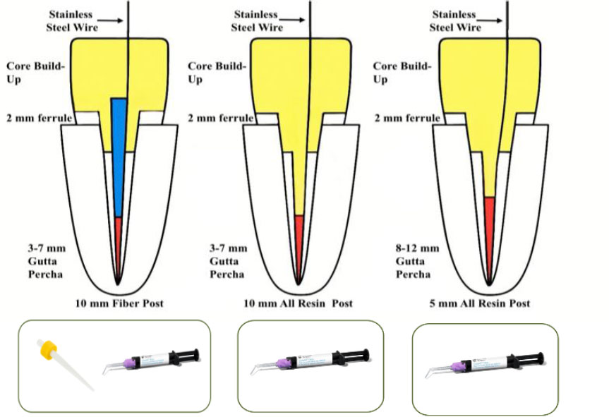
Illustrates three post-and-core designs differing in post material, length, and remaining gutta-percha, all incorporating a stainless steel wire and a 2 mm ferrule, to enable comparative evaluation of their biomechanical performance.
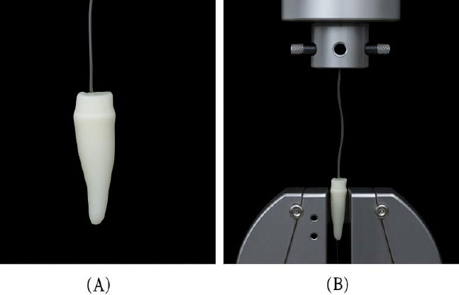
(A) Image showing the experimental setup of a specimen suspended by wire prior to mechanical testing, (B) Image showing the specimen positioned within the universal testing machine.
2.3. Mechanical Testing Procedure
Retention strength was assessed using a universal testing machine (Instron 5944, Canton, MA, USA). Each specimen was positioned vertically, and the embedded stainless-steel orthodontic wire was secured to the upper holding unit of the device (Fig. 2B).
A vertical tensile force was applied at a crosshead speed of 0.5 mm/s until failure occurred, which was confirmed by an audible signal from the machine and separation of the post-and-core assembly. The maximum force at failure was recorded in Newtons (N).
All tests were performed under controlled laboratory conditions. To minimize variability and operator bias, standardized procedures were followed by trained personnel. A flowchart summarizing the experimental steps is provided in Fig. (3).
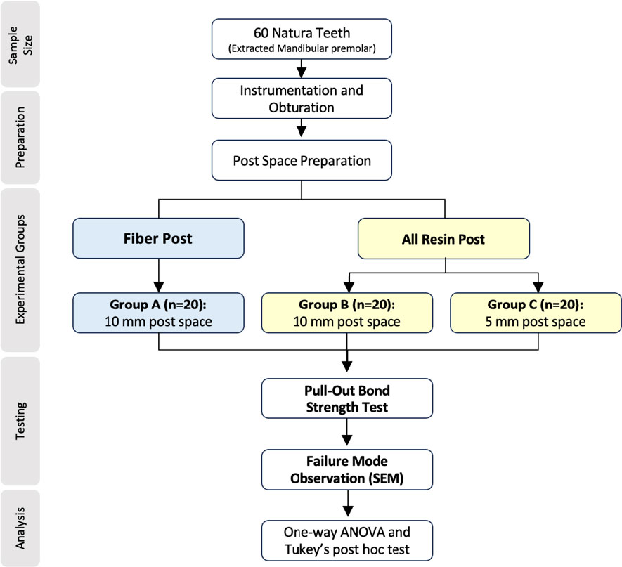
Flowchart illustrating the experimental design.
2.4. Instrumentation and Measurement
This investigation employed a universal testing machine (Instron 5944) (Fig. 4), calibrated before testing to ensure accurate measurement of tensile forces up to 2 kilonewtons (kN). This calibration confirmed data stability and enabled precise assessment of retention strength across the different post configurations.
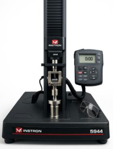
Testing of specimens was performed using a universal testing machine (Instron 5944, Norwood, MA, USA) equipped with a 2 kN load cell to conduct pull-out tests for evaluating the retention strength of the tested materials.
The dependent variable was retention strength, evaluated with the pull-out test, while the independent variables were post type and post length. To minimize operator bias, all procedures were standardized, random allocation was used for group assignments, and all measurements were performed by trained personnel to maintain consistency and reliability.
2.5. Statistical Analysis
Data were processed and analyzed using SPSS version 26 (IBM, USA). Because the dataset met the assumptions for parametric analysis, a one-way Analysis of Variance (ANOVA) was applied to compare mean values of pull-out strength among the three groups.
When significant differences were detected, Tukey’s Honestly Significant Difference (HSD) test was used for post-hoc pairwise comparisons to identify specific intergroup differences. ANOVA was chosen because it is appropriate for comparing more than two independent groups with normally distributed data. A p-value less than 0.05 was considered statistically significant.
3. RESULTS
Sixty samples were evaluated and categorized into three groups according to post type and length. Pull-out bond strength was measured for each group, and the mean retention strength was calculated.
3.1. Pull-out Bond Strength
Group A (10 mm fiber post) exhibited a mean pull-out bond strength of 942.9 ± 41.38 N. Group B (10 mm resin-based post) demonstrated the highest mean value of 978.6 ± 56.63 N, while Group C (5 mm resin-based post) showed the lowest mean value of 758.6 ± 73.82 N. These findings are summarized in (Table 1).
| Group | Mean Pull-out Strength (N) | Standard Deviation (SD) | 95% Confidence Interval (N) |
| Group A | 942.9 | 41.38 | (917.92, 967.93) |
| Group B | 978.6 | 56.63 | (944.4, 1012.83) |
| Group C | 758.6 | 73.82 | (714.01, 803.22) |
A one-way Analysis of Variance (ANOVA) revealed a statistically significant difference in retention strength among the groups (p = 0.024). Tukey’s Honestly Significant Difference (HSD) post-hoc test showed no significant difference between Groups A and B (p > 0.05). However, significant differences were found between Groups A and C and between Groups B and C (p < 0.05). These findings are summarized in Fig. (5).
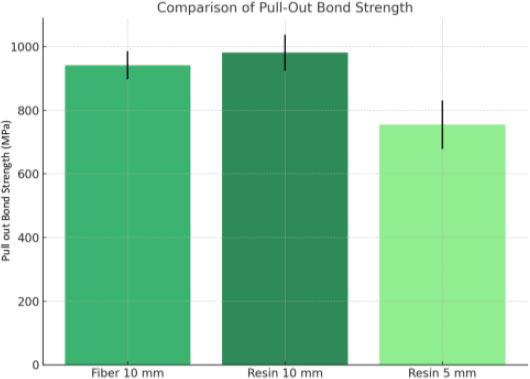
Bar chart showing the mean pull-out bond strength (N) for the three study groups.
3.2. Failure Mode Analysis
Following pull-out testing, all samples were examined under a microscope to determine failure modes.
-
Groups B and C (10 mm and 5 mm resin-based posts, respectively) showed predominantly favorable failures, defined as debonding at the cement–post interface without structural damage to the root (Fig. 6A).
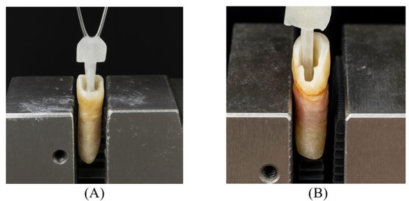 Fig. (6).
(A) Post debonding, whereas (B) root fractures extending into the cementoenamel junction and are considered unfavorable.
- Group A (10 mm fiber post) exhibited a higher proportion of unfavorable failures, characterized by root dentin fractures that rendered the specimens non-repairable (Fig. 6B).
Scanning Electron Microscopy (SEM) revealed three distinct failure modes across all groups:
-
Adhesive failure: exposed dentinal tubules indicating debonding at the dentin–resin interface (Fig. 7A).
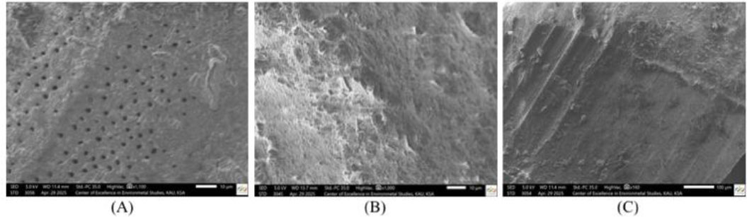 Fig. (7).
SEM micrographs illustrating failure modes: (A) Adhesive failure with visible dentinal tubules, indicating debonding at the resin-dentin zone; (B) Cohesive failure limited to the resin layer; (C) Mixed failure exhibiting characteristics of both adhesive and cohesive types.
- Cohesive failure: fractured resin remaining on both substrates, indicating internal failure within the resin material (Fig. 7B).
- Mixed failure: features of both adhesive and cohesive failure, with areas of exposed dentin and fractured resin remnants (Fig. 7C).
3.2. Summary
Overall, the 10 mm resin-based posts (Group B) had the highest mean pull-out bond strength, followed by the 10 mm fiber posts (Group A), while the 5 mm resin-based posts (Group C) had the lowest strength among the tested groups. Significant differences were observed between Groups B and C, and between Groups A and C, as presented in Fig. (8) as a box plot of retention strength. The predominant and secondary failure modes identified after pull-out testing for each experimental group are summarized in (Table 2).
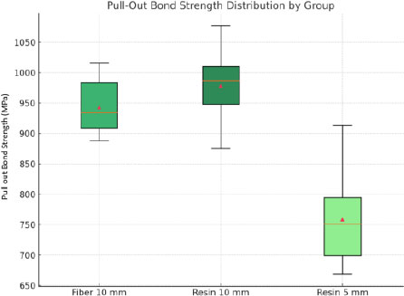
Box plot of pull-out bond strength distribution for the three study groups. Group B (10 mm resin-based post) and Group A (10 mm fiber post) showed higher median and mean bond strengths than Group C (5 mm resin-based post), which exhibited the lowest values and the greatest variability.
| Group | Predominant Failure Mode | Secondary Failure Modes | Overall Failure Nature |
|---|---|---|---|
| Group A (Fiber post – 10 mm) | Root dentin fracture | Adhesive, mixed | More unfavorable |
| Group B (Resin-based post – 10 mm) | Adhesive debonding | Cohesive, mixed | Predominantly favorable |
| Group C (Resin-based post – 5 mm) | Adhesive debonding | Mixed | Predominantly favorable |
4. DISCUSSION
In the rehabilitation of Root Canal–Treated (RCT) teeth, post-and-core systems are frequently required to restore structural integrity and facilitate restorative procedures [25, 3]. Considering the high prevalence of RCTs and posts in durable outcomes, this topic remains clinically relevant [14]. Despite the variety of available post materials, including fiber, metal, and resin-based, there is no consensus on the optimal material and length [26, 12]. Previous studies have examined material properties and retention, yet the relationship between post length and bonding capacity, particularly when comparing resin-based and fiber posts, is not fully resolved [1]. This study addresses the gap by comparing resin-based and fiber posts at two different lengths (10 mm vs 5 mm).
A 5 mm post length was selected based on clinical guidelines suggesting that post length should be equal to or slightly greater than the clinical crown height to ensure proper retention and resistance form. Based on an average clinical crown (with ferrule and ceramic clearance) of approximately 4–5 mm, a 5 mm post length is considered the minimal acceptable threshold of post length when used in constrained anatomical cases [27]. Although resistance form was not directly tested in this study, pull-out bond strength was used as a surrogate indication for post retention under tensile loading conditions. The novelty lies in evaluating material and length under controlled in vitro conditions to isolate their effects on pull-out bond strength. Our methodology followed established principles: pull-out testing with a universal testing machine (Instron) provides consistent force application approximating clinical mechanical stresses [28], and the in vitro model allowed systematic control over different variables [29]. Furthermore, the use of human premolars enhances the clinical applicability [30].
The 10 mm resin-based posts exhibited the highest pull-out bond strength, followed by fiber posts with 10 mm length, while 5 mm resin-based posts showed the lowest retention. These findings reflect the increased bonded surface area associated with longer posts, supporting adhesive principles and aligning with prior studies stating that long posts enhance mechanical retention [30, 31]. To increase the bonded surface area without weakening the root, the remaining dentin thickness should be maintained at least 1 mm, and the preparation should not exceed a third of the root diameter [32]. This supported by a recent FEA finding that indicated maintaining 1 mm thickness of radicular dentin is more critical than post material [33]. Sun et al. further established that excessive canal enlargement leads to more stress in the cervical area and post-apex, thereby increasing the fracture risk [34].
Fiber posts have slightly lower values compared to resin-based posts of the same length; this may be attributed to material-specific bonding behavior. Fiber posts provide better stress distribution due to their dentin-like elastic modulus [25], but the resin-based posts benefit from the chemical continuity with the resin cement to form direct resin-to-resin bonding [9, 12, 26]. Due to their favorable biomechanical performance, fiber post systems are widely used due to their favorable biomechanical properties, but their bonding performance under various clinical conditions has not been questioned [35]. Boschian Pest et al. (2002) observed reduced bond strengths of fiber posts when chemical adhesion to dentin was limited, emphasizing the importance of achieving strong adhesive interfaces in post retention [24]. Despite their marginally lower retention, fiber posts remain clinically useful in reducing catastrophic root fractures through favorable stress distribution [8, 25]. The sharp drop in the retention observed in the 5 mm resin-based group reinforces the critical influence of post length, which is consistent with previous findings [1, 5, 11].
Pull-out bond strength is considered an important indicator for post retention, but it does not necessarily predict the mode of failure. Therefore, evaluating failure mode in addition to retention becomes the key factor for post-system evaluation [15]. The failure mode is significantly affected by the post space diameter more than the length, as shown in previous studies. Increased post length may result in increased fracture risk when the remaining dentin thickness is not preserved [36, 37].
Failure mode analysis revealed adhesive, cohesive, and mixed failures across all groups, consistent with previous studies using similar pull-out or push-out testing methods [5, 28, 38]. Group A (10 mm fiber) exhibited a slightly higher frequency of unfavorable failures, including fractures extending to the cemento-enamel junction, although no statistically significant differences in failure distribution were detected among groups. Favorable or repairable failures were most frequent in shorter post groups, possibly due to lower retention values and reduced stress concentration within root dentin [11, 14]. These findings align with previous evidence that post geometry, bonding effectiveness, and restorative integrity collectively have an impact on failure behavior [8, 25, 31]. In contrast, Marinescu et al. reported that deeper fiber posts placed into the root canal (7 mm) have slightly higher fracture resistance than shorter lengths (5 mm) [39]. Potential differences between the studies may arise from different methodologies and different factors affecting the failure mode, like the post material. Overall, despite differences in retention values, the tested groups produced comparable failure patterns under in vitro loading conditions.
Within the limits of this study, both post length and material influenced retention outcomes. The superior pull-out performance of 10 mm resin-based posts suggests that, when adequate length is achievable, they may provide strong bonding due to direct resin-to-resin continuity [12, 26]. Nevertheless, fiber posts remain the preferred clinical option in many situations because of their favorable stress distribution and lower risk of catastrophic root fractures [8, 25, 31]. Clinicians should therefore consider both retention capacity and fracture behavior when selecting post systems, prioritizing fiber posts in cases where preservation of root integrity is essential, while reserving resin-based posts for situations where maximum bonding capacity can be achieved without compromising root structure.
As an in vitro investigation, this study does not fully replicate the complexities of the clinical environment. Important variables such as thermal cycling, fatigue loading, and occlusal stresses were not simulated, which may influence long-term bonding and fracture outcomes. Only single-rooted premolars were tested, limiting the generalizability of results to teeth with more complex anatomies. In addition, a single pull-out protocol was applied, whereas different loading modes may produce variable results [5, 28, 38]. Future studies should incorporate cyclic fatigue, thermal aging, and dynamic loading to approximate clinical conditions more closely, as well as assess the impact of cement film thickness, alternative post geometries, and different cementation techniques [15, 31].
CONCLUSION
Within the limitations of this in vitro study, post length was found to significantly influence pull-out bond strength in endodontically treated premolars. The 10 mm resin-based posts exhibited retention comparable to that of 10 mm fiber posts, suggesting that they can serve as a conservative alternative to prefabricated fiber posts. Shorter 5 mm resin-based posts exhibited reduced retention, suggesting that they may be insufficient in cases where high mechanical stability is required. Regarding failure mode, fiber posts showed a higher frequency of unfavorable failure modes compared with all-resin posts. These findings reinforce the importance of optimizing post length to enhance retention while minimizing sound radicular dentin removal. Future clinical studies are recommended to validate these in vitro results under functional and aging conditions.
AUTHORS’ CONTRIBUTIONS
The authors confirm their contribution to the paper as follows: M.A, A.A, O.A.: Study conception and design; R.A, R.Al, O.A, W.A.: Data collection; M.A, A.A, O.A.: Analysis and interpretation of results; M.A, R.A, R.A.l.: Draft manuscript preparation. All authors reviewed the results and approved the final version of the manuscript.
LIST OF ABBREVIATIONS
| RCT | = Root Canal–Treated |
| SEM | = Scanning Electron Microscopy |
| HSD | = Honestly Significant Difference |
| FRC | = Fiber-Reinforced Composite |
ETHICS APPROVAL AND CONSENT TO PARTICIPATE
This study was conducted in Jeddah, Saudi Arabia, after receiving ethical approval from the Research Ethical Committee at the Faculty of Dentistry, King Abdulaziz University (KAUFD) (Approval No. 124-11-24).
HUMAN AND ANIMAL RIGHTS
This experimental in vitro study was conducted in accordance with the principles of the Declaration of Helsinki.
CONSENT FOR PUBLICATION
This study did not involve personal data, images, or audio-video materials from individual participants. Extracted teeth were obtained with informed consent from donors prior to use, and no identifying information was collected or published.
AVAILABILITY OF DATA AND MATERIALS
The data and supportive information are available within the article.
ACKNOWLEDGEMENTS
The authors would like to thank the Advanced Technology Dental Research Laboratory, Faculty of Dentistry, King Abdulaziz University, Jeddah, Saudi Arabia, for supporting this experiment.